MCI Snippets: New PET/CT scanner improves imaging tests
New technology at the Mitchell Cancer Institute enables physicians to capture better images of patients with PET and CT scans, making it easier to detect cancer.
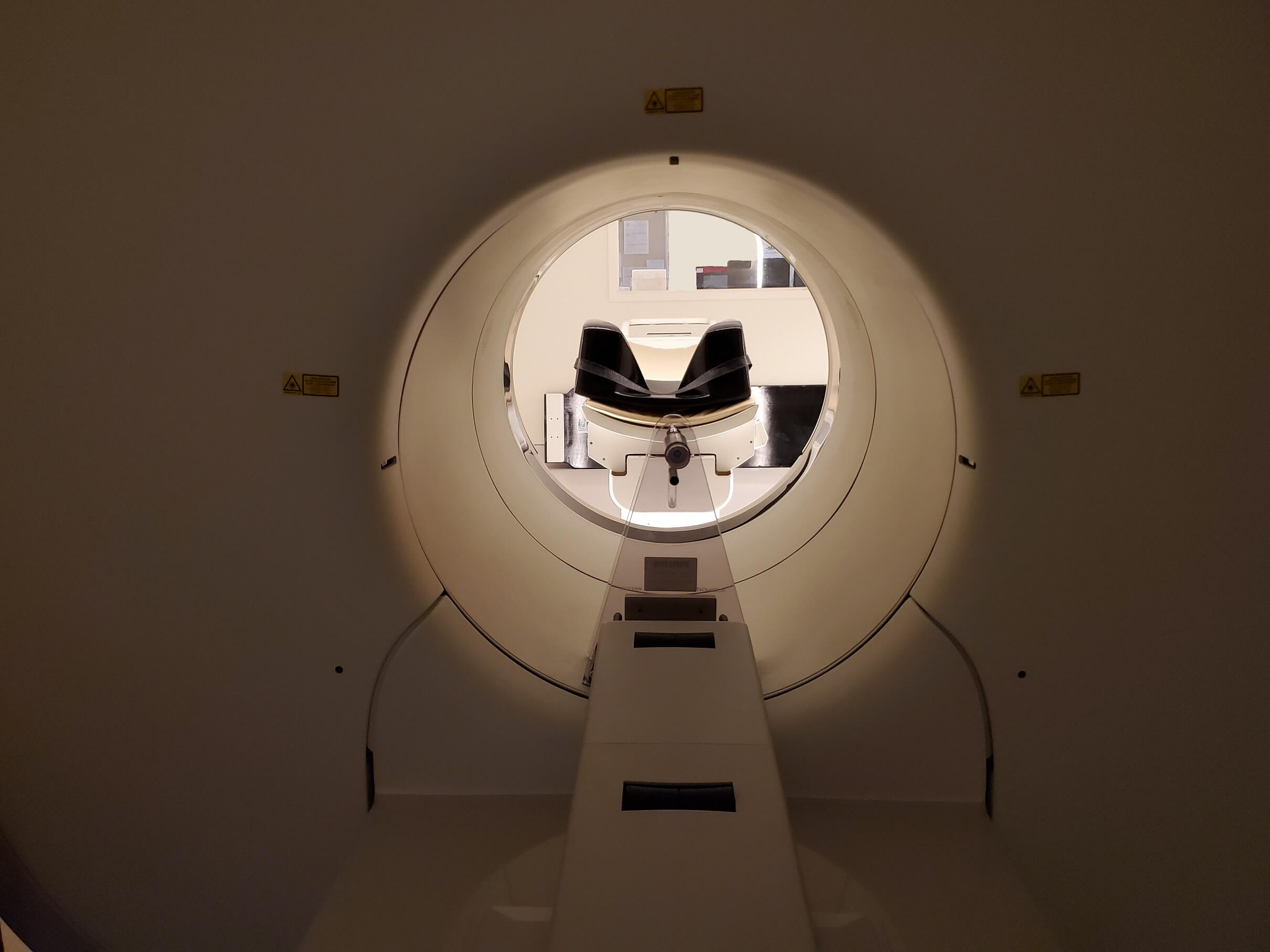
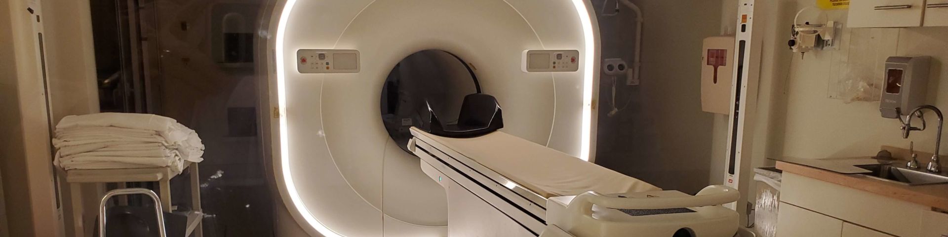
The new Philips Vereos digital PET/CT scanner, installed recently at the main Mobile office, takes the MCI’s scanning capability from analog to digital. Shikha Gupta, M.D., M.P.H., the primary radiologist for the MCI and professor of radiology at the USA College of Medicine, explains it this way: “A digital clock displays the exact time in numbers, while the analog clock relies on telling time with hands,” she said. “Analog is approximate. Digital is specific.”
In an oncology setting, digital scanning improves the quality of the images compared with those obtained by analog scanners. The backbone of the new scanner is a digital photon-counting detector that uses solid-state sensors to count the individual scintillation photons created during a PET scan, Gupta said. By contrast, analog scanner detectors merely record flashes of light.
Gupta said that the new technology also can shorten scan times, making the process easier and more comfortable for patients. “The ability to detect smaller lesions and characterize them with greater confidence is particularly useful in lung nodules, head and neck, and breast cancer,” she said. “This improvement in image quality is even more significant in overweight or obese patients.”